Article Text

Statistics from Altmetric.com
Bacteria of the species Chlamydia trachomatis are divided into serovars that are associated with different disease manifestations. Serovars A-C cause trachoma, which occurs mainly in undeveloped countries. Serovars D-K are responsible for oculogenital infections, and serovars L1, L2, and L3 cause lymphogranuloma venereum (LGV). Infections of serovars A-K are usually confined to the mucosal epithelia of the eyes and the anogenital tract. In contrast, the L-serovars are more invasive and may induce genital ulcer or inguinal lymphadenopathy after passing the epithelial surface.1
While serovars D-K are distributed worldwide and represent the most frequent bacterial sexually transmitted disease in Europe and North America, LGV caused by the L-serovars is a very rare disease in industrialised countries, but is restricted to parts of southeast Asia, Africa, South America, and the Caribbean.2
During the second part of 2003 three patients presented to our clinic with inguinal swellings. In addition, genital ulcer developed in two of them. All patients had homosexual contacts with more than one partner. Two patients were HIV positive, one of them refused HIV testing. The patients assured us they had not travelled outside Germany during the past year.
In all cases genital C trachomatis infection was diagnosed by DNA amplification in lesional swabs or lymph node aspirates using the SDA technology (ProbeTec ET, Becton-Dickinson, MD). Other infections inducing genital lesions were not detected. None of the patients had a positive serology indicating active infection with Treponema pallidum. Genital infections due to Neisseria gonorrhoeae, Haemophilus ducreyi, and herpes simplex virus were excluded by polymerase chain reaction (PCR) testing. In addition, no genital bacterial and fungal pathogens were detectable by direct microscopy or culture.
After treatment with doxycycline (200 mg per day), genital lesions completely regressed in all patients. Patients 2 and 3 were treated for 3 weeks, while patient 1 received doxycycline for 1 week only.
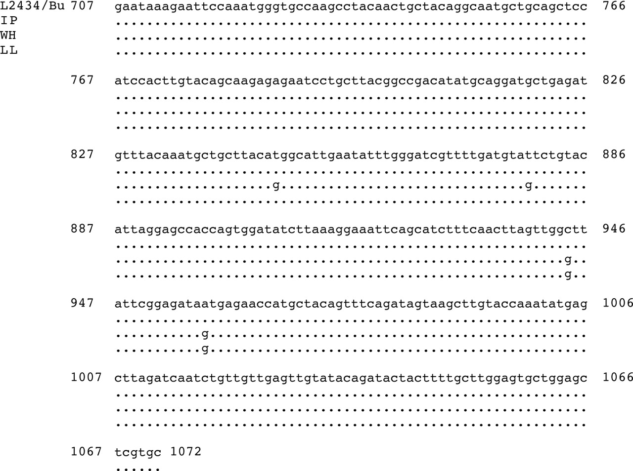
The underlying C trachomatis serovars were identified by sequence analysis of ompA derived DNA fragments amplified using primers MF21 and MB22 as described by Dean et al.3 In all cases the sequences obtained had highest homology to C trachomatis serovar L2 but were not identical. While the sequence from patient 1 was shown to be 100% identical to the L2 isolate 434/Bu4 over the analysed region of 366 nucleotides, the sequences of patients 2 and 3 were only 89.9% and 99.5% homologous, indicating different sources of infection (fig 1).

DNA sequence alignment of MOMP-PCR fragments. Nucleotide numbers are according to C trachomatis L2 strain 434/Bu (Genbank Acc No M14738). IP, WH, and LL are the initials of the patients who were C trachomatis positive. Homology to 434/Bu is 100% (IP), 98.9% (WH), and 99.5% (LL), respectively.
Recently, a cluster of 15 LGV cases among homosexual men was reported in Rotterdam.5 Thirteen of these patients were HIV positive. As with our patients, C trachomatis serovar L2 was identified in all patients. Although anonymous sexual contacts in Germany were reported, there is yet no epidemiological evidence for a connection of the LGV cases of our study and those reported in Rotterdam.
In conclusion, infections with C trachomatis serovar L may be more frequent than assumed previously, as indicated by the identification of three different strains in our study. Consequently, LGV should be included in the differential diagnosis of genital ulcer or lymphadenopathy in homosexual HIV infected patients in Europe.
Linked Articles
- Brief Encounters