Article Text

Abstract
Objectives: To describe a series of lymphogranuloma venereum (LGV) cases presenting as inguinal syndrome and/or genital ulceration seen among men who have sex with men (MSM) in London, UK.
Methods: Collaborative retrospective case note review. Clinicians from three London genitourinary medicine (GUM) clinics accessed by large populations of MSM within the current LGV outbreak collected clinical data from confirmed cases of LGV inguinal syndrome or genital ulcer. LGV was confirmed by the detection of LGV-specific DNA from specimens such as bubo aspirates, ulcer swabs, urethral swabs, first void urine and rectal biopsy material.
Results: There were 13 cases detected overall: 5 cases of urethral LGV infection with inguinal adenopathy, 3 cases of genital ulcer with LGV inguinal adenopathy, 3 cases of isolated LGV-associated inguinal buboes, 1 case of a solitary LGV penile ulcer and 1 case with a penile ulcer and bubonulus. Only 6 of the 13 were HIV positive and all tested negative for hepatitis C. The majority of cases reported few sexual contacts in the 3 months preceding their diagnosis.
Conclusions: Clinical manifestations of LGV in MSM have not been confined to proctitis in the current outbreak in the UK and a small but significant number of inguinogenital cases of LGV have been observed. Epidemiologically, many of the cases described seem to have occurred at the periphery of the current MSM LGV epidemic. Clinicians need to be vigilant for these less common presentations of LGV among MSM and specific diagnostic tests should be done from the relevant lesions.
Statistics from Altmetric.com
Lymphogranuloma venereum (LGV) is caused by Chlamydia trachomatis serovars L1–L3.1 These serovars display tropism for the lymphatic system in contrast to serovars A–K that affect mucocutaneous tissue.1 Endemic in Africa, the Caribbean and parts of Asia,1 the classical presentation of LGV is inguinal lymphadenitis and abscess formation sometimes preceded by anogenital ulceration. Since 2004 there has been a resurgence of LGV proctitis affecting predominantly men who have sex with men (MSM) who are HIV positive in the UK, Western Europe and the USA.2–4 Detected cases have had high rates of co-infection with other sexually transmitted infections (STIs), including hepatitis C.2 Anogenital ulceration and/or lymphadenitis has been reported rarely in the current MSM LGV epidemic.2 5
We describe 13 recent cases of LGV that presented with “classical” inguinogenital manifestations. LGV was detected, usually in the absence of other STIs, and symptoms resolved after treatment with long-course regimens of doxycycline or azithromycin.
METHODS
Clinicians from three inner London genitourinary medicine (GUM) clinics servicing large populations of MSM collected clinical data from confirmed cases of LGV inguinal syndrome or genital ulcer. Case histories were anonymised and relevant data were collated and compared. Written informed consent was obtained from all cases that provided clinical images for this paper. As this was a retrospective anonymised case note review, ethics committee approval was not sought from the participating centres.
Initial tests for C trachomatis and other STIs were performed at the respective hospital laboratories using commercially available assays performed according to the instructions of the manufacturer. All 13 cases described had specimens confirmed as LGV using molecular tests performed at the Sexually Transmitted Bacteria Reference Laboratory (STBRL), Health Protection Agency, Colindale, UK. DNA was extracted from clinical specimens (ulcer swabs, urethral swabs, first void urine) using the MagNA Pure total NA kit (Roche Diagnostics GmbH, Mannheim, Germany) according to the manufacturer’s instructions. In instances where limited clinical specimen was available (bubo aspirates and rectal biopsies), manual DNA extractions were performed using the QIAamp Viral Mini Kit (Qiagen, Crawley, UK). The chlamydia status of all referred specimens was confirmed initially using a plasmid-based real-time PCR method.6 The presence of LGV DNA was then detected using a LGV-specific pmpH real-time PCR assay.7
RESULTS
Clinical records
The characteristics of the 13 cases are summarised in table 1.
All were MSM with a median age of 36 years (range 21–61). Ten were of white ethnicity, only six were infected with HIV and none tested positive for hepatitis C antibodies. Twelve cases presented with unilateral or bilateral inguinal adenopathy, four of whom had concurrent genital ulceration, and one patient presented with penile ulceration only. Five of the twelve men with inguinal adenopathy appear to have had urethral infection as their primary LGV lesion and none of the five reported any preceding anogenital ulceration, although such symptoms might have been transient and unnoticed. Six of the men reported only solitary sexual contacts in the previous 3 months, three of whom were their HIV positive regular male partners.
Notable features of cases
Of note, case 1 was first seen in December 2003 when he developed a tender right inguinal swelling. He reported regular unprotected insertive and receptive anal sex with anonymous male partners in London in the preceding months. On examination there was a single fluctuant 4 cm by 5 cm right inguinal swelling. Fine needle aspiration biopsy showed reactive lymphoid hyperplasia; Gram stain, microscopy and culture of the aspirate were negative for bacterial pathogens including mycobacteria. Oral amoxicillin 500 mg three times daily and flucloxacillin 500 mg four times daily were prescribed with no improvement in the mass, which ruptured spontaneously and discharged pus periodically for several months. The patient then began to pass red blood per rectum, developing tenesmus and showing weight loss. Colonoscopy in October 2004 showed an “unusual, chronic-looking fissure” in the anal canal and a purulent proctitis, biopsies of which showed acute and chronic inflammation with crypt abscesses and focal granuloma formation, consistent with LGV. A rectal swab specimen (ProbeTec, Becton Dickinson, Sparks, Maryland, USA) tested positive for C trachomatis by strand displacement amplification (SDA). The inguinal and rectal symptoms resolved completely after 3 weeks of doxycycline treatment. LGV-specific DNA was detected retrospectively from rectal biopsy material using techniques described previously.8
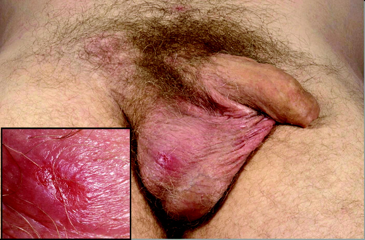
Case 2 also had a complicated clinical course after first presenting in September 2005 with a week’s history of a 5 mm tender ulcer over the right hemiscrotum and a healing perianal ulcer. He was prescribed a course of aciclovir and flucloxacillin but a swab from the ulcer tested negative for both herpes simplex virus (HSV) and C trachomatis (ulcer swab and urethra). The patient re-presented 2 weeks later with worsening of the ulcer that was now indurated with granulation tissue evident in the base (see fig 1). This time an ulcer swab specimen tested positive for C trachomatis by SDA subsequently confirmed to be LGV. Five days after testing the patient was recalled and by this time had developed an enlarged firm right inguinal node and was thus commenced on doxycycline 100 mg twice daily for presumed LGV. After 10 days he had developed further lymphadenopathy above the inguinal ligament despite compliance with treatment. After 17 days of doxycycline, the swelling had progressed to a hot, fluctuant, 4 cm by 3 cm bubo confirmed with ultrasound. Treatment was switched to azithromycin 1 g daily but despite the change in antibiotic treatment the bubo continued to enlarge. Surgeons were reluctant to incise the lesion due to extensive overlying cellulitis but the abscess ruptured spontaneously 2 weeks after starting azithromycin. A sample of fluid draining from the sinus also tested positive for C trachomatis and was later confirmed to be LGV. Symptoms improved on azithromycin, reduced to 500 mg daily for the last 5 days. The lesions showed complete resolution after a total of 5 weeks treatment (18 days azithromycin and 17 days doxycycline) though some residual skin induration remained.

Clinical images from case 2 showing a small tender indurated scrotal ulcer (inset) and early right-sided inguinal lymphadenopathy that progressed to 3 cm by 4 cm bubo formation prior to spontaneous rupture. Lymphogranuloma venereum DNA was isolated from both ulcer swab and bubo pus.
Case 4 had a 2-month history of a solitary, weeping, painless, indurated 0.7 cm ulcer in the dorsal coronal sulcus yet never developed inguinal lymphadenopathy.
Case 6 presented with bilateral 2 cm by 3 cm fluctuant lymph node abscesses that had ruptured spontaneously. The aspirated pus tested positive for C trachomatis by SDA and confirmed to be LGV.
Case 7 presented with striking clinical signs of a left-sided 12 cm by 6 cm inguinal mass and a smaller 4 cm by 2 cm mass on the right. An ultrasound study arranged by his general practitioner had shown multiple enlarged pathological-appearing lymph nodes bilaterally. A urethral swab specimen tested positive for C trachomatis by chlamydial cell culture and Roche Cobas Amplicor PCR (Roche Diagnostics Systems, Branchburg, New Jersey, USA) and extracts from both were confirmed to be LGV. The lesions resolved completely without rupture after a 3 week course of doxycycline.
Case 8 was admitted to hospital for investigation of bilateral inguinal lymphadenopathy with a differential diagnosis including lymphoma. His urine had tested positive for C trachomatis by SDA 4 days before and he was reviewed in the GUM clinic where a diagnostic aspirate was performed from the non-fluctuant lymph node mass using 0.5 ml normal saline (see fig 2) and the blood-stained material obtained was deposited onto a ProbeTec female swab. C trachomatis was detected using SDA and was confirmed to be LGV as was the DNA extract from his initial chlamydia-positive urine specimen. His symptoms and lesions resolved after 3 weeks of doxycycline treatment.

Clinical images from case 8 showing massive right inguinal lymphadenopathy that was tender but non-fluctuant. Inset shows aspiration of the mass with 0.5 ml normal saline using a lateral approach, which obtained some blood-stained fluid that tested positive for lymphogranuloma venereum DNA.
Case 9 also had a diagnostic aspirate performed from non-fluctuant inguinal lymphadenopathy and was started on doxycycline treatment. He returned the following day after the mass had doubled in size to 8 cm by 4 cm but it was still non-fluctuant. Urgent ultrasound assessment showed a cluster of enlarged lymph nodes up to 3 cm each in size with surrounding cellulitis and oedema but only a small 1.4 cm by 0.5 cm abscess situated deeply and not amenable to drainage. Doxycycline treatment was continued for 3 weeks and the symptoms and mass resolved without further suppuration.
Case 11 presented with a non-tender, non-fluctuant, lymph node swelling, yet 1 ml of frank pus was aspirated from the node and this tested positive for C trachomatis and confirmed to be LGV (fig 3).

Clinical images from case 11 demonstrating right-sided non-tender, non-fluctuant lymphadenopathy. Despite this, 1 ml of frank pus (see inset) was aspirated from the node and this tested positive for lymphogranuloma venereum DNA.
Case 12 presented with a 3 cm by 5 cm right inguinal lymph node mass and asymptomatic LGV urethritis yet the non-purulent lymph node aspirate tested negative for C trachomatis by SDA.
Case 13 presented with a penile ulcer and subsequently developed unilateral inguinal lymphadenopathy as well as a penile bubonulus—only the second case to be described in the recent MSM LGV epidemic.9 His new male partner was asymptomatic but had LGV detected from a rectal swab specimen.
DISCUSSION
The cases described in this series differ epidemiologically from the typical MSM LGV proctitis cases seen thus far in the UK epidemic. Less than half was HIV positive compared with 74% of proctitis cases2 and other concurrent STIs were not detected. Half of the cases appear to have contracted LGV from relatively isolated episodes of sexual risk with far fewer recent sexual contacts than reported from most proctitis cases, particularly those seen early in the epidemic.2 Notably, most of the present cases reported no downstream sexual contacts following the onset of their symptoms and thus were unlikely to have transmitted LGV to subsequent partners. The prompt symptomatology and management seen in cases 2–13 suggests that they are unlikely to have been a source of onward transmission of LGV. In case 13, the index case’s asymptomatic partner was diagnosed subsequently with rectal LGV, reminding us that asymptomatic LGV exists in the MSM population. This is in accordance with findings from the Netherlands where 40% of men with LGV proctitis reported few complaints and/or had no physical abnormality.10 Nevertheless, a recent UK case finding exercise failed to demonstrate a significant reservoir of asymptomatic infection to explain persistent transmission11 and further work is needed to define true differences in LGV epidemiology between Dutch and British MSM.
While patient 1 was not diagnosed with LGV inguinal syndrome contemporaneously, his inclusion in the present series serves to demonstrate the consequences of missed diagnosis. He presented in an era when LGV was not recognised as a prevalent pathogen in the UK MSM population and much morbidity and onward transmission could have been averted by early diagnosis and treatment. It is not possible to determine if his eventual LGV proctitis was due to progression of his inguinal syndrome or from newly acquired anorectal infection.
Diagnosis of LGV is dependent on the detection of a LGV-associated serovar of C trachomatis from the site of pathology; however, serology may be helpful should this fail and the clinical suspicion is high. Only three of five cases in our series showed typical serological responses and more work is needed to assess the diagnostic value of serology in this population.
The LGV genital ulcers seen in this series showed non-specific clinical features and clinicians should consider obtaining suitable specimens for C trachomatis/LGV when assessing anogenital ulceration in MSM. Swabs from ulcers and diagnostic aspirates of scant material from non-fluctuant inguinal lesions produced suitable specimens for the successful detection and typing of C trachomatis using standard nucleic acid amplification assays and we recommend this approach for investigation of suspicious lesions.
Urethral C trachomatis appears to have been the primary LGV lesion in five of the present cases. Variability in symptomatology and urethral smear microscopy findings in the present cases suggest that this might not simply represent LGV-associated urethritis and that other primary endourethral lesions such as ulceration may occur. We do not believe that routine LGV typing of C trachomatis-positive urethral isolates from MSM is currently indicated, based on the rarity of urethral LGV seen in the UK case-finding exercise (Ward H, in press). Nevertheless, in the presence of additional clinical signs such as ulceration or lymphadenopathy, or in LGV contacts, then referral of C trachomatis-positive specimens for detection of LGV serovars is warranted.
The presence of persistent anogenital ulceration and severe proctitis can enhance HIV12 and possibly hepatitis C transmission.13 Although no new diagnoses were made within the present case series, incident HIV and hepatitis C infection may not have been detected by testing performed at the time of LGV diagnosis and follow-up serology beyond the relevant window periods is indicated.
Treatment with doxycycline for 3 weeks achieved resolution of symptoms and signs in 11 of 12 patients and azithromycin in 2 patients using multiple-dose regimens. As demonstrated in patient 2, diagnosis of LGV can be challenging and patients with large buboes may require longer courses of treatment than the recommended 3 weeks.14 Adjuvant drainage of fluctuant abscesses may hasten the resolution of such lesions and prevent spontaneous rupture and sinus formation. There were no signs of tertiary sequelae such as chronic lymphoedema reported in any of the present cases although some local scarring was seen.
The present cases demonstrate that clinical manifestations of LGV have not been confined to proctitis in the current outbreak in MSM in the UK despite the predominance of anorectal disease reported. Clinicians who see MSM patients should familiarise themselves with the clinical features and diagnostic pathways illustrated in this series. The striking clinical signs seen in some of these cases, especially in those who were systemically unwell, led to provisional diagnoses of incarcerated herniae and lymphoma. In addition, there has been a recent report of two heterosexual LGV cases:15 further evidence that suggests the epidemic is spreading beyond its initial core group. Other relevant clinicians, including surgeons, microbiologists and histopathologists, should be alerted to the current epidemiology of LGV in the UK, Europe and the USA, and it should be considered in the differential diagnoses of proctitis, anogenital ulceration and inguinal lymphadenopathy, particularly in MSM.
Key messages
-
Genital ulcers and inguinal node disease caused by lymphogranuloma venereum (LGV) have been observed in the current LGV epidemic in London in men who have sex with men (MSM).
-
Chlamydia trachomatis nucleic acid tests performed on swabs, urine and lymph node aspirates provide suitable diagnostic specimens that allow confirmation by LGV-specific molecular analysis.
-
LGV should be included in the differential diagnosis of anogenital ulceration in all MSM.
Acknowledgments
The authors would like to thank the laboratory staff at St Thomas’ Hospital, Mortimer Market Centre, and Chelsea and Westminster for their assistance in analysing specimens; Health Protection Agency Bristol for performing the chlamydial serology in case 4; and Professor Cathy Ison for helpful comments on the manuscript.
REFERENCES
Footnotes
-
See Editorial, p 157
-
Competing interests: None.
-
Patient consent: Obtained.
-
Contributors: GS and JW conceived the paper, collected data on cases seen at St Thomas’ Hospital, co-ordinated the multicentre collaboration and wrote the final draft; EA-J, JR, NTA and DH each contributed cases from their respective clinics and edited the manuscript; AE wrote the early first draft; SA was responsible for the molecular typing and verification of LGV results as well as editing the manuscript.
Linked Articles
- Editorial
- Correction