Article Text

Abstract
Objectives Pharyngeal Chlamydia trachomatis (chlamydia) might contribute to ongoing chlamydia transmission, yet data on spontaneous clearance duration are rare. We examined the prevalence, spontaneous clearance, chlamydial DNA concentration and genotypes of pharyngeal chlamydia among clinic patients with sexually transmitted infection (STI).
Methods Female patients at high risk for an STI who reported active oral sex and male patients who have sex with men (MSM) were screened for pharyngeal chlamydia RNA using a nucleic acid amplification test. A repeat swab was obtained to evaluate spontaneous clearance in untreated patients with pharyngeal chlamydia. Quantitative chlamydia DNA load was determined by calculating the chlamydia/human cell ratio.
Results Pharyngeal chlamydia was detected in 148/13 111 (1.1%) MSM and in 160/6915 (2.3%) women. 53% of MSM and 32% of women with pharyngeal chlamydia did not have a concurrent anogenital chlamydia infection. In 16/43 (37%) MSM and in 20/55 (36%) women, the repeat pharyngeal swab was negative (median follow-up 10 days, range 4–58 days). Patients with an initial chlamydial DNA concentration above the median were less likely to clear. Of 23 MSM with pharyngeal chlamydia who had sex with a lymphogranuloma venereum (LGV)-positive partner recently or in the past, two were LGV biovar positive (8.7%).
Conclusions The pharynx is a reservoir for chlamydia and LGV, and may play a role in ongoing transmission. Although delay in ribosomal RNA decline after resolution of the infection might have led to an underestimation of spontaneous clearance, in high-risk STI clinic patients, testing the pharynx for chlamydia should be considered.
- CHLAMYDIA TRACHOMATIS
- ORAL CAVITY
- ORAL SEX
- SCREENING
- TESTING
Statistics from Altmetric.com
Introduction
Urogenital infection with Chlamydia trachomatis (CT) is the most prevalent bacterial sexually transmitted infection (STI) in most industrialised countries, including the Netherlands.1 Lymphogranuloma venereum (LGV), a serious ulcerative STI caused by C. trachomatis biovar L, is endemic among men who have sex with men (MSM).2 Although treatment is straightforward, STI screening programmes have been unable to stop the ongoing epidemics of both C. trachomatis and LGV.3
Dutch STI guidelines recommend screening for pharyngeal C. trachomatis (PhCT) on indication only4 and the United States Centers for Disease Control does not recommend testing for PhCT.5 The British Association for Sexual Health and HIV recommends PhCT screening of MSM and commercial sex workers reporting sexual risk behaviour that may result in pharyngeal infection.6
The relevance of PhCT for the individual patient and for ongoing chlamydia transmission is unknown. Testing for PhCT in various STI clinic populations using a nucleic acid amplification test (NAAT) revealed a PhCT prevalence between 1% and 3%.7 ,8 Although one study showed an association between upper respiratory tract symptoms and PhCT,9 most patients with PhCT are asymptomatic.8 ,10 ,11 Recently, acquisition of urethral chlamydia through fellatio has been reported in heterosexual males and MSM.12 ,13 Depending on the duration of pharyngeal infection, PhCT might play an important role in the ongoing transmission of chlamydia.
In this study, we investigated pharyngeal C. trachomatis RNA prevalence, spontaneous clearance and bacterial DNA load in a large group of MSM and women visiting the STI clinic in Amsterdam, The Netherlands. We also performed LGV genotyping on PhCT samples from a subset of MSM to examine whether the pharynx is a potential reservoir for LGV.
Methods
Study setting
The STI outpatient clinic at the Public Health Service of Amsterdam (GGD Amsterdam) annually performs around 36 000 free-of-charge and anonymous STI consultations. All patients are routinely tested for C. trachomatis, gonorrhoea, syphilis, hepatitis B14 and also for HIV using an opt-out strategy.15 Routinely collected data (eg, age, country of birth, number and type of sexual contacts in the previous 6 months, history of HIV, symptoms, laboratory results, diagnoses and spontaneously reported pharyngeal symptoms) are registered in an electronic patient database, are also registered.
In this article, four substudies are reported: (1) C. trachomatis prevalence among those routinely tested for PhCT, (2) spontaneous clearance of PhCT, (3) chlamydial DNA concentration in PhCT samples and (4) LGV typing in PhCT samples from MSM at increased risk for LGV. Inclusion of subjects was between January 2011 and July 2012.
Substudy 1: prevalence of and predictors for pharyngeal chlamydia
At our clinic, women are regarded as having a high-risk profile if they report STI-related symptoms, if they were notified by a sexual partner with an STI or if they are involved in commercial sex. High-risk women who report active fellatio in the previous 6 months (regardless of condoms use) and all MSM (irrespective of reported active fellatio) are routinely tested for pharyngeal Neisseria gonorrhoeae and C. trachomatis. This test is performed on a nurse-collected pharyngeal swab using the APTIMA Combo 2 assay (GEN-PROBE, San Diego, California, USA). For self-collected urine samples (MSM), nurse-collected cervical swabs (women) and nurse-collected rectal samples (from those reporting passive anal sex in the previous 6 months) C. trachomatis is tested using the APTIMA CT assay (GEN-PROBE). Approximately 1 week after the initial consultation, definitive results are available and patients who need treatment are invited to come to the clinic at the shortest notice for a follow-up visit and treatment; the date of treatment consultation is determined by the patient. In case of PhCT, a single dose of azithromycin 1000 mg is offered, unless azithromycin or doxycycline (100 mg twice daily for a minimum of 7 days) was already prescribed as presumptive treatment at the initial consultation.
Substudy 2: spontaneous clearance of pharyngeal chlamydia
PhCT-positive patients who returned for treatment, who were over 18 years of age and did not take antibiotics at their first visit were asked for written informed consent to participate in the substudy on spontaneous clearance of pharyngeal chlamydia. During the first 5 months, only patients with PhCT and without concurrent anogenital C. trachomatis, anogenital gonorrhoea and pharyngeal gonorrhoea infection were invited to participate. When the diagnosis became available, eligible visitors consenting to participate were asked to have pharyngeal swabs collected three more times (when the diagnosis was communicated, plus 1 and 2 weeks later), whereafter treatment was given. However, due to the high number of patients with a coinfection and the low proportion of patients who agreed to participate, we decided to change the inclusion criteria (approved by the ethical committee), and henceforth (remaining 13 months) all patients with PhCT were invited to participate. In the new design, upon inclusion, after the diagnosis became available, a nurse collected a single follow-up pharyngeal swab (APTIMA Combo 2 assay) and, using a questionnaire, interviewed the patient about oral sexual behaviour between the initial and follow-up visits. Participants were subsequently treated for PhCT as described above.
Substudy 3: quantification of chlamydial DNA concentration
In all chlamydia-positive pharyngeal samples from initial and follow-up consultations, quantitative chlamydia bacterial load determination was done, using a real-time PCR targeting the cryptic plasmid (expressed as inclusion-forming units (IFU)). In the same samples, the number of human leucocyte antigen (HLA) copies was determined using a quantified serial dilution. The chlamydia/human cell ratio (chlamydial DNA concentration) was calculated as described previously16 and expressed as IFU/100 million HLA copies. As the majority of PhCT samples tested negative in the less sensitive real-time PCR, analysis on a linear scale had very limited power. As a solution, samples were categorised in three groups: (1) chlamydial DNA undetectable (C. trachomatis-positive in the APTIMA Combo 2 assay but negative in the real-time PCR), (2) chlamydial DNA concentration equal or below and (3) chlamydial DNA concentration above the median. Samples in which no HLA was detected were excluded from this analysis.
Substudy 4: testing for pharyngeal LGV
Pharyngeal samples from MSM with PhCT who also had (1) a concurrent diagnosis of anorectal or inguinal LGV or (2) a history of LGV, or (3) a history of sexual contact with a partner with LGV, were selected for pharyngeal C. trachomatis biovar L testing. PhCT samples from initial consultation were tested with a pmpH-based in-house real-time PCR to discriminate between LGV and non-LGV genotypes, as described previously.17 ,18 If both the LGV and non-LGV test results were negative in the pmpH test, the result was considered inconclusive.
Statistical analysis and data collection
All statistical analyses were performed using SPSS V.19 (SPSS, Chicago, Illinois, USA) and STATA Intercooled V.11.0 (STATA, College Station, Texas, USA). Sexual preference and commercial sex work refers to the period 6 months prior to the consultation. Ethnicity was defined based on criteria of Statistics Netherlands (CBS)19 and was categorised in Dutch and non-Dutch. HIV status was based on the HIV test result at the initial consultation or on self-reported HIV-positive status. A concurrent diagnosis of STI was defined as being diagnosed with chlamydia at another anatomical location, gonorrhoea, infectious hepatitis B, HIV and/or infectious syphilis at the initial consultation. Anorectal chlamydia status (only tested when reporting receptive anal sex) was categorised in three groups (not tested, negative and positive).
χ2 Test or Fisher's exact test was used to compare categorical variables between groups; the Mann–Whitney U test was used to compare continuous variables between groups. p Values of <0.05 were considered statistically significant. Univariate and multivariable logistic regression analyses were conducted to identify independent determinants of PhCT. Multivariable model building was done using a backward step-wise procedure, including only those variables with a univariate p value of <0.10. Age and number of sexual partners in the previous 6 months were forced into and kept in the model. Other variables were kept in multivariable models if p<0.05. Due to small numbers, multivariable analyses of determinants for PhCT spontaneous clearance could not be performed.
Results
Prevalence of and predictors for pharyngeal chlamydia
Between January 2011 and July 2012, 48 653 consultations were performed at the STI clinic, of which 13 339 with MSM and 8078 with high-risk women (38.5% of all female patients). At 13 111 and 6915 consultations, respectively, pharyngeal swabs were tested for chlamydia (figure 1).

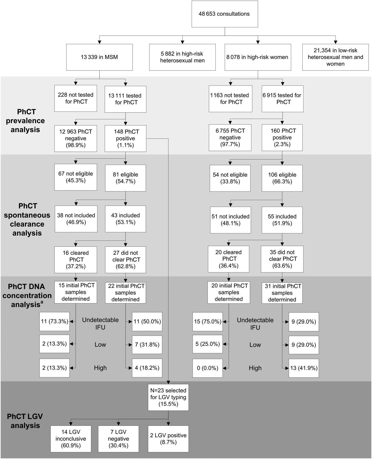
Flow chart of pharyngeal Chlamydia trachomatis (PhCT) sub-studies: (1) prevalence of PhCT; (2) spontaneous clearance of pharyngeal C. trachomatis; (3) analysis of C. trachomatis DNA concentration in pharyngeal infections; (4) prevalence of pharyngeal LGV, STI clinic, Public Health Service of Amsterdam, 2011–2012. HLA, human leucocyte antigen; IFU, inclusion forming units; LGV, lymphogranuloma venereum; MSM, men who have sex with men; PhCT, pharyngeal C. trachomatis; STI, sexually transmitted infection. aChlamydial DNA concentration defined as; low: equal or lower than median (3.4 log IFU/100 million HLA copies); high: higher than median (3.4 log IFU/100 million HLA copies); undetectable IFU: IFU undetectable.
The PhCT prevalence in MSM was 1.1% (148 diagnoses; 95% CI 0.9 to 1.3, table 1).
Associations of baseline characteristics with pharyngeal chlamydia in 13 111 men who have sex with men (MSM), STI clinic, Public Health Service of Amsterdam, 2011–2012
Two MSM with PhCT did not return to the clinic to obtain their treatment. Pharyngeal symptoms were reported by 285 (2.2%) PhCT-negative MSM and by 2 (1.4%) PhCT-positive MSM (p=0.49). Concurrent anogenital chlamydia infections were found in 1280 (9.9%) PhCT-negative MSM and in 70 (47.3%) PhCT-positive MSM (p<0.001). In multivariable analyses, PhCT positivity was associated with being notified of any STI, concurrent anorectal chlamydia, concurrent urogenital chlamydia, more than 10 sexual partners in the previous 6 months, and an unknown HIV status (versus HIV-negative).
PhCT prevalence among women was 2.3% (160 diagnoses; 95% CI 2.0 to 2.7, table 2).
Associations of baseline characteristics with pharyngeal chlamydia in 6915 high-risk women, STI clinic, Public Health Service of Amsterdam, 2011–2012
Four women with PhCT did not return to the clinic to obtain their treatment. Pharyngeal symptoms were reported by 26 (0.4%) PhCT-negative women and 1 (0.6%) PhCT-positive woman (p=0.47). Concurrent anogenital chlamydia infections were found in 658 (9.7%) PhCT-negative and in 109 (68.1%) PhCT-positive women (p<0.001). In multivariable analyses, women reporting commercial sex work had a lower risk for PhCT, while those with pharyngeal gonorrhoea, those notified of an STI, those with a concurrent urogenital chlamydia and those with more than 10 sexual partners in the previous 6 months had a higher risk for PhCT.
Spontaneous clearance of pharyngeal chlamydia
Out of 148 MSM and 160 women with PhCT, 81 (54.7%) and 106 (66.3%) met the inclusion criteria, respectively (figure 1). Not showing up for treatment, not willing to participate and mistakenly not being invited resulted in 43 (53.1%) MSM and 55 (51.9%) women with PhCT participating in the follow-up study.
MSM who participated were significantly less often notified of an STI (16.3% vs 34.3%; p=0.028), had less often STI-related symptoms (14.0% vs 37.1%; p=0.005) and had less often an anogenital chlamydia infection (32.6% vs 53.3%; p=0.022). Women who participated were significantly older (median age 24 vs 22 years; p=0.005), received more often payment for sex in the previous 6 months (37.0% vs 17.0%; p=0.005), were less often notified of an STI (25.9% vs 49.1%; p=0.005) and had less often a concurrent urogenital chlamydia infection (55.6% vs 74.5%; p=0.015).
The median time between initial and follow-up consultation was 12 days for MSM (range, 7–58 days) and 9 days for women (range, 4–58 days). At follow-up, PhCT was cleared in 16/43 (37.2%) MSM and in 20/55 (36.4%) women (see online supplementary table S1). Half of MSM and 46.3% of women reported active fellatio without a condom since initial consultation, but this did not affect spontaneous clearance (p=1.0 in MSM and p=0.88 in women). Median follow-up time in MSM and women who cleared PhCT (13.0 (IQR 9–19) and 8.5 (IQR 8–13.5)) did not significantly differ from those who did not clear (10.0 (IQR 8–14), p=0.15; and 9 (IQR 8–15), p=0.67, respectively). No significant determinants for PhCT spontaneous clearance were detected among MSM (see online supplementary table S2), but among women, increasing age was associated with spontaneous clearance (per-year increase in age: OR 1.13; 95% CI 1.01 to 1.27); see online supplementary table S3). Concurrent urogenital chlamydia infection was inversely associated with spontaneous clearance (OR 0.32; 95% CI 0.10 to 1.00).
Quantification of chlamydial DNA concentration
In 88 (91.7%) out of 96 participants of the clearance substudy with available samples, HLA could be detected in both (initial and follow-up) PhCT samples (figure 1). From the 88 initial samples, 46 (52.3%) had an undetectable, 23 (26.1%) a low and 19 (21.6%) a high chlamydial DNA concentration (table 3).
Chlamydial DNA concentration in initial and follow-up pharyngeal Chlamydia trachomatis positive samples and association with spontaneous clearance of pharyngeal chlamydia in 88 patients, STI clinic, Public Health Service of Amsterdam, 2011–2012
Among the 46 participants with an undetectable chlamydial DNA concentration, 26 (56.5%) had cleared the infection, compared with only 7 (30.4%) and 2 (10.5%) among those with low and high chlamydial DNA concentrations (p=0.001). No consistent pattern was observed in the chlamydial DNA concentration in follow-up samples compared with the initial samples (table 3).
Lymphogranuloma venereum
Totally, 23 MSM with PhCT at initial consultation had a current or past LGV diagnosis or reported sexual contact with a partner with LGV (figure 1). Out of 23 pharyngeal samples, two were LGV biovar positive (8.7%; these were not included in the bacterial clearance substudy), 7 (30.4%) negative and 14 (60.9%) inconclusive.
Discussion
Principal findings
In comparison with urogenital and anorectal C. trachomatis, the prevalence of pharyngeal C. trachomatis is lower among MSM and high-risk women tested at this large STI clinic. Nonetheless, 52.7% and 31.9% of MSM and female patients with PhCT did not have a concurrent anogenital C. trachomatis infection; they would not have been treated if screening had only been performed for anogenital C. trachomatis infections. During follow-up, a minority (36.7%) of patients spontaneously cleared PhCT. Those with a higher chlamydial DNA concentration were less likely to spontaneously clear PhCT.
Strengths and weaknesses
A strength of this study is the high number of MSM and women screened for PhCT. To date, this study reports the largest number of MSM and women screened for PhCT at both initial and follow-up consultations. A limitation of this study was that less than half the patients with PhCT had a follow-up consultation at which they were retested.
Whereas C. trachomatis DNA can be positive in the presence of remnants from non-viable organisms, the presence of C. trachomatis RNA implies that bacterial replication occurs and is, therefore, seen as an indicator of potential infectious bacterial viability.20 Although C. trachomatis cultivation in follow-up samples would definitely prove bacterial persistence, the sensitivity of C. trachomatis cultivation is too low to be considered in clinical studies. Testing for chlamydial ribosomal RNA after azithromycin treatment in women with urogenital chlamydia showed that after 10 days 34% were still rRNA positive.21 This delay in rRNA decline after resolution of the infection might have led to an underestimation of the proportion with spontaneous PhCT clearance in our study.21 ,22 Also, it is unknown whether pharyngeal swabs are the preferred method to detect PhCT. Instead of pharyngeal swabs, the use of oral wash specimens (whereby a larger surface of the oral mucosa can be sampled) might be considered for future studies.23
The real-time PCR used for the quantification of C. trachomatis load is less sensitive than the APTIMA Combo 2 assay.24 ,25 In this study, the majority of PhCT samples were negative in the real-time PCR implying a low level of chlamydial DNA in these samples.
Relation to other studies
In San Francisco, sentinel surveillance among MSM in 2010 showed a PhCT prevalence of 1.7%, slightly higher than the 1.1% found in our study.7 In a Dutch study among female STI clinic patients reporting fellatio (January 2007–July 2008), PhCT prevalence was similar to that among high-risk women in our study (1.9% vs 2.3%).8
In another report from San Francisco in 2003, PhCT without concurrent anogenital C. trachomatis infections (solitary PhCT) was found in 30 out of 50 MSM (60.0%),26 comparable with the 52.7% found in our study. In the Dutch study among women, the proportion of women with solitary PhCT was similar to that among high-risk women in our study (31% vs 31.9%).8
A relative small proportion of MSM and women spontaneously cleared PhCT during follow-up. In two earlier studies with a limited number of PhCT-positive participants, in 1 out of 2 (50%) cases with a median follow-up of 11 days27 and in 6 out of 18 (33.3%) with a mean follow-up of 8 days (SD±6), PhCT was cleared during follow-up.23
Meaning of the study findings
This study shows that the pharynx is a potential reservoir for both chlamydia and LGV. Oral-to-genital transfer of C. trachomatis might be possible since PhCT RNA is detectable in a majority of patients for more than 1–2 weeks. Therefore, screening for PhCT may be relevant from a public health point of view. In this study, more than 93.0% of MSM and 85.6% of high-risk women reported fellatio in the previous 6 months, of whom, only 1.5% and 9.2% always used a condom during oral sex, respectively. The high frequency of unprotected orogenital contact in combination with the observed low spontaneous clearance might result in onward transmission of chlamydia.
Participants with an undetectable chlamydial DNA concentration at initial consultation more often cleared their PhCT, suggesting that the bacterial load present in the pharynx determines spontaneous clearance. Participants with PhCT but with an undetectable bacterial load at initial consultation more often cleared the infection, suggesting that the bacterial load influences risk of spontaneous clearance. Patients with PhCT and with an undetectable chlamydial DNA load may have less impact on the ongoing transmission, as in many of them C. trachomatis resolves within a short time. However, patients with a high chlamydial DNA load carry the bacteria for a longer period and likely transmit it via fellatio.
Strong predictors for PhCT in both women and MSM were concurrent urogenital chlamydia, being notified of an STI by a sexual partner, and high numbers of sexual partners. Among MSM, anorectal chlamydia was also significantly associated with PhCT. These predictors can be explained by increased risk for pharyngeal exposure to a chlamydia-positive partner. Unknown HIV status was a predictor for PhCT in MSM. These MSM actively declined being tested for HIV; they are known to exhibit higher sexual risk behaviour.15 Female sex workers were less likely to have PhCT. This can be explained by more consistent and professional condom use. Women with pharyngeal gonorrhoea were at higher risk for PhCT. Possibly these women are at high risk for having sexual contact with a chlamydia-infected partner or pharyngeal gonorrhoea might increase the susceptibility for PhCT. Spontaneous reported pharyngeal symptoms were rare in patients with PhCT. Consequently, there are no symptom-based indications to test for PhCT, and testing only those patients who are at higher risk for PhCT will result in missed cases.
Unanswered questions and future research
To date, no large analysis has been performed to estimate the risk of oral-to-genital transfer of C. trachomatis. Our study focused on high-risk women and MSM. The observed prevalence and clearance may differ from those in other female and heterosexual male patients at the STI clinic. Since PhCT might contribute to the urogenital chlamydia epidemic, cost-effectiveness studies are needed to justify routine PhCT screening.
In conclusion, PhCT is found in 1.1% of MSM and 2.3% of high-risk women visiting the STI clinic in Amsterdam, The Netherlands. A minority of the PhCT patients cleared PhCT RNA within 1–2 weeks, and a lower C. trachomatis DNA concentration was associated with spontaneously cleared PhCT. PhCT might be a reservoir for the transmission of chlamydia and LGV.
Key messages
After a median follow-up of 10 days, only 37% of patients with pharyngeal Chlamydia trachomatis had cleared their infection.
Clearance was associated with a lower chlamydial DNA concentration.
32% (women) and 53% (men who have sex with men) of cases with pharyngeal chlamydia did not have concurrent anogenital chlamydia infections.
Acknowledgments
The authors would like to thank all the nurses working on this project and all the participants of this study. Special thanks goes to Titia Heijman and Antoinette van Roosmalen for their assistance in planning the study, to Martine Schlüter for the logistics at the laboratory, and to Jannie van der Helm for her suggestions on the chlamydial DNA concentration analysis. The authors would also like to thank Jolein Pleijster (Technician, VUmc) for technical assistance in bacterial load determination.
References
Supplementary materials
Supplementary Data
This web only file has been produced by the BMJ Publishing Group from an electronic file supplied by the author(s) and has not been edited for content.
Files in this Data Supplement:
- Data supplement 1 - Online supplement
Abstract in Dutch
This web only file has been produced by the BMJ Publishing Group from an electronic file supplied by the author(s) and has not been edited for content.
Files in this Data Supplement:
- Abstract in Dutch - Online abstract
Footnotes
Previously presented: Information from this paper has been orally presented at the NVED (1 February 2013, Lunteren, The Netherlands) and at the STI & AIDS World Congress (17 July 2013, Vienna, Austria; abstract number O18.6).
Handling editor Jackie A Cassell
Contributors MSvR, MFSvdL and HJCdV designed the study protocol, supported by APvD and AGCLS. MSvR was responsible for implementation and data collection at the STI clinic. APvD and AGCLS were responsible for the chlamydia and LGV diagnostics at the laboratory. SAM was responsible for the chlamydial DNA load determination. MSvR and MFSvdL performed the statistical analyses. MSvR, MFSvdL and HJCdV drafted the paper, all authors commented on draft versions, and all approved the final version.
Funding This work was supported by the Research and Development Fund of the Public Health Service of Amsterdam, grant no. 2137.
Competing interests None.
Ethics approval This study was approved by the Medical Ethical Committee of the Academic Medical Centre, University of Amsterdam, Amsterdam, The Netherlands (ethics approval number MEC 10/216 # 11.17.416).
Provenance and peer review Not commissioned; externally peer reviewed.